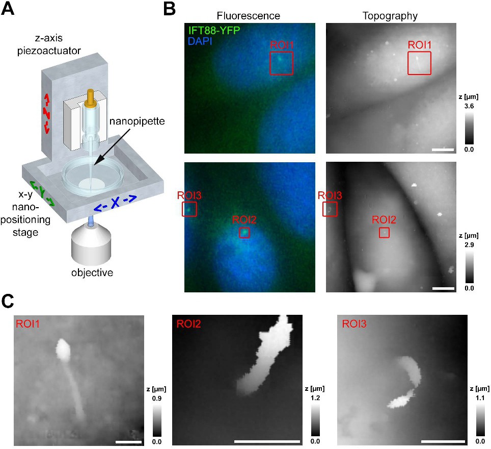
Fig. 1. Identification of primary cilia in IMCD IFT88-YFP epithelial cells using hopping mode SICM and epifluorescence. Schematic of the SICM setup mounted on top of epifluorescence microscope (A). Epifluorescence (B, left) and height-coded SICM topography images (B, right) of the same area of fixed (4% paraformaldehyde) kidney epithelial cells showing the fluorescent cilia in green (IFT88 labelled with YFP) and the nucleus with DAPI (1µg/ml). High-resolution topography of primary cilia (C) identified in the individual regions of interest ROI1-ROI3 marked in (B). Scale bars 5 µm (B), 1 µm (C).